Projects
Subject areas
Correlative X-ray Imaging (CXI) is continuously evolving, developing and integrating new methodologies and optical components over the years, with contributions from multiple researchers, for example.
Spatial-Spectral, Time-Resolved Multi-Contrast X-ray and NMR
This research aims to establish a comprehensive framework for data acquisition and analysis in multicontrast imaging, offering enhanced spatial, spectral, and temporal resolution. The proposed approach integrates advanced techniques, including Image Processing Software, Two-Dimensional and Three-Dimensional Hybrid and Correlative Imaging, as well as Computer Vision algorithms.
Size-Selective X-ray Imaging
Implementation of single-shot X-ray imaging for advanced characterization of structures at the nanometric scale. Beam modification induced by the optical element facilitates three contrast modalities: transmission, differential phase contrast, and small-angle scattering. This approach enables the analysis of nanostructures without the need for direct resolution.
Customized technological development for multi-contrast X-ray imaging
Hartmann masks are developed using UV lithography and gold electroforming, along with Shack–Hartmann-type wavefront sensors (SHXS) produced via 3D-DLW in the form of two-dimensional lens arrays. By combining SHXS with super-resolution techniques, the analysis of multi-contrast images is significantly improved.
Synthesis and chemical characterization of polymers
Development of negative-tone epoxy resins for lithography. In collaboration with EMPA, Switzerland, Aerosol Jet Printing was used to fabricate 3D pillar arrays from photopolymeric inks. Polypy framework was created to interpret polymer properties from mass Spectroscopy Data.
Optical element fabrication – X-ray Hartmann mask
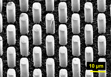
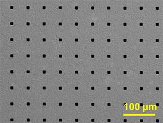
A number of applications require time-resolved measurements of dynamic processes at different time scale. In such approaches, versatile and robust single-shot X-ray imaging methods are preferable, such as non-interferometric X-ray imaging techniques with a single optical element in the beam path. Such element introduces periodic beam modulation and could be, for instance, represented by two-dimensional array of microstructures, e.g. Hartmann masks (absorbing grid) or newly developed inverted Hartmann masks (an array of absorbing pillars). In this project, we develop, characterize and employ customized transmission X-ray optics for single-shot X-ray imaging within tabletop X-ray radiography setup. Manufacturing of customized Hartmann masks is performed using widely accessible UV lithography technique and gold electroplating. To ensure highest photon efficiency, we utilize low-absorbing substrates such as graphite and polyimide with conductive coatings.
 |
 |
|
| An array of gold pillars – inverted Hartmann mask | Golden grid – conventional Hartmann mask |
Method development – Size-selective X-ray imaging
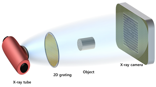
In single-shot X-ray imaging method, the beam modulation introduced by the optical element is distorted by the presence of the sample in the beam path. Analysis of these distortions yields three contrast modalities: transmission, differential phase contrast and small-angle scattering. An intrinsic property of the signal associated with small-angle scattering has dragged a lot of attention because it can serve as a window in the world of nanostructures in the real space without directly resolving them. The scattering image in this case is the auto-correlation of the electron density distribution at a specific distance. This gives an opportunity to probe different scattering lengths by changing the setup arrangement. Single-shot imaging setup due to its versatility and robustness offers a wide range of possible adjustments to achieve a desirable scattering length scale. The use of two-dimensional optical components such as Hartmann mask, allows scattering into two dimensions to be detected, making the method sensitive to sample anisotropy. Due to enhanced sensitivity to electron density proposed method can lead to better understanding of structure-property relationships. The proposed technique is promising for various materials science applications involving low-absorbing materials with internal nanostructures (e.g. functional polymer nanocomposites).
Synthesis and chemical characterization of an X-ray sensitive polymer material for microstructure patterning
The main objective is to synthesize and characterize a customized photoresist for X-ray lithography application.
Using qualitative and quantitative characterization methods, we are trying to understand the lack of reproducibility within the X-ray lithography process. The polymer synthesis is studied in a way to vary the chain sizes, which will end up in the same material but with different mechanical properties. The evaluation of different formulations for the photoresists is also a current activity. When the optimal formulation is developed and the process parameters adjusted, a characterization methodology as quality control of the photoresist will be standardized.
Activities: polymerization synthesis, spectroscopy characterization methods, formulation of photoresists, µ-LIGA fabrication, and image/quality control techniques such a contrast curve, visibility and homogeneity maps.
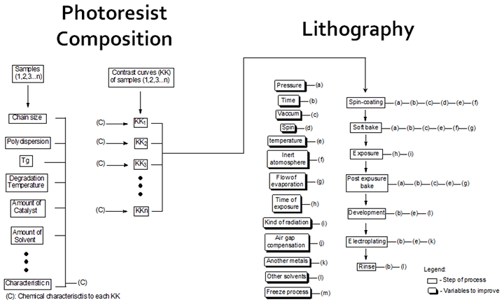
Scheme of the photoresist composition characterization methods, lithography process steps, and its parameters.
Technology development for emerging multi-contrast X-ray imaging using a cone beam configuration
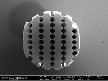
In the past decades several phase-sensitive methods were proposed to overcome the limitations of conventional attenuation-based radiography to investigate low density materials or with similar absorption cross section. Among these techniques, phase-sensitive imaging by means of grating interferometry is widely employed to enhance the contrast of weakly absorbing materials. Differential phase-contrast configuration differs from the standard radiography setup by the addition of several optical elements, gratings, between the X-ray source and the detector. We propose to develop a novel non-destructive method to analyze wavefront distortions induced by low-absorbing materials in the hard X-ray imaging regime. The method is based on multi-contrast X-ray imaging by means of a single exposure, and replacing the several gratings elements arrangement in interferometric setups with a single three-dimensional photoresist grating structure (Fig. 1) in the beam path.
 |
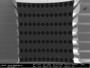 |
Fig1. 3D grating-like structures made by 3D 2-photon lithography
Currently, I devote time to the design and manufacturing of large-scale three-dimensional optical elements to be employed as Shack-Hartmann sensors for investigation of dynamical processes. Furthermore, within my Ph.D. thesis we are developing an X-ray optics laboratory at the IMT.

